Anatomy of the Distal Humerus
The distal humerus is divided into the medial column and the lateral column.
Medial Column
The medial column includes the medial part of the metaphysis of the humerus, the medial epicondyle, and the medial condyle, including the trochlea of the humerus.
Lateral Column
The lateral column includes the lateral part of the metaphysis of the humerus, the lateral epicondyle, and the lateral condyle, including the capitulum of the humerus.
Coronoid Fossa and Olecranon Fossa
Located between the two columns are the anterior coronoid fossa and the posterior olecranon fossa.
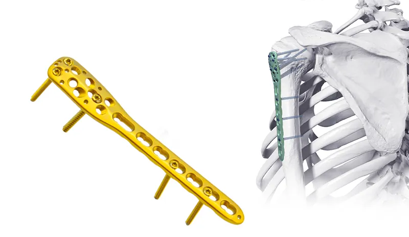
Distal Humeral Extraarticular Locking Plate
This plate is designed for extraarticular fractures of the distal humerus, targeting the metaphyseal region without involving the joint surface. Its locking screws provide angular stability, making it particularly suitable for osteoporotic patients or complex fracture patterns.
Extraarticular Fixation Multi-directional Locking Screws Anatomic Contouring Combination Holes
Mechanism of Injury in Distal Humerus Fractures
The distal humerus is divided into the medial column and the lateral column.
Why is the distal humerus prone to fractures?
As a crucial part of the elbow joint, distal humerus fractures often result from "direct trauma" (such as a fall landing on the elbow) or "indirect trauma" (such as twisting or throwing actions).
- Muscle Pulling ForcesLateral Column
The medial column includes the medial part of the metaphysis of the humerus, the medial epicondyle, and the medial condyle, including the trochlea of the humerus. ·Strong contraction of internal rotator muscles ·Strong contraction of elbow flexor muscles
- High-Energy Trauma
External forces such as traffic accidents or falls from height can result in comminuted fractures or involve the articular surface.
Coronoid Fossa and Olecranon Fossa
·Traffic accidents ·Falls from height
Distal Lateral Humeral Locking Plate
Designed for lateral column fractures, this plate is suitable for capitellar or supracondylar fractures, providing mechanical support via a lateral approach to facilitate early rehabilitation.
Lateral Column Stabilization Articular Fragment Fixation Radial Nerve Protection Dynamic Compression
AO Classification: Accurate Typing to Guide Surgical Decisions
According to the AO classification system, distal humerus fractures are categorized as follows, directly influencing the choice of treatment strategies:
Type A: Extra-articular fractures
- A1: Simple fracture, no articular involvement. - A2/A3: Impacted or displaced metaphyseal fractures, attention needed for angular deformities.
Type B: Partial articular fractures
Fracture line involves the joint surface but without complete separation; anatomical reduction is necessary to restore joint congruency.
Type C: Complete articular fractures
- C1/C2: Simple intra-articular fractures with metaphyseal involvement. - C3: Comminuted intra-articular fractures, the most challenging to treat; stable fixation is needed to reduce the risk of post-traumatic arthritis.. Loc
Significance of Classification:
The AO classification clarifies the severity of the fracture and provides guidance for plate selection (e.g., single vs. dual plates, locking screw designs) to ensure both fixation strength and biological balance.
Distal Medial Humeral Locking Plate
This plate is an internal fixation solution for medial column fractures of the distal humerus, providing support via a medial approach to prevent varus deformity, often used in cases with coronal plane fractures.
Medial Column Support Low-profile Design Coronal Plane Screws Dual-plating Compatible
Surgical Treatment: Core Advantages of Plate Fixation
Treatment Principles:
Following the AO philosophy: "Anatomical reduction, stable fixation, and early functional exercise."
High-Energy Trauma
External forces such as traffic accidents or falls from height can result in comminuted fractures or involve the articular surface.
Treatment Principles
Surgical Indications
Articular displacement >2mm
Open fractures
Combined neurovascular injury
Failure of conservative treatment
Plate Fixation Strategie
Suitable for type C fractures. Fixation from both medial (e.g., anatomical locking plate) and lateral (e.g., parallel plate) sides provides 3D stability and reduces the risk of postoperative rotational deformity.
Used for type A and partial type B fractures. Pre-contoured plates conforming to distal humerus anatomy minimize soft tissue dissection.
Combined with percutaneous screw placement to reduce infection risk and preserve periosteal blood supply.
Clinical Value of Plate Fixation
Biomechanical Advantage
Locking plates provide angular stability, especially beneficial for osteoporotic patients.
Functional Recovery Guarantee
Anatomical reduction preserves elbow joint mobility to the greatest extent, reducing complications such as nonunion or malunion.
Customized Design
Plates shaped for specific fracture types (e.g., intercondylar ridge support plates) optimize force transmission and accelerate bone healing.
Anatomy of the Distal Humerus
The distal humerus is divided into the medial column and the lateral column.
Medial Column
The medial column includes the medial part of the metaphysis of the humerus, the medial epicondyle, and the medial condyle, including the trochlea of the humerus.
Lateral Column
The lateral column includes the lateral part of the metaphysis of the humerus, the lateral epicondyle, and the lateral condyle, including the capitulum of the humerus.
Coronoid Fossa and Olecranon Fossa
Located between the two columns are the anterior coronoid fossa and the posterior olecranon fossa.