


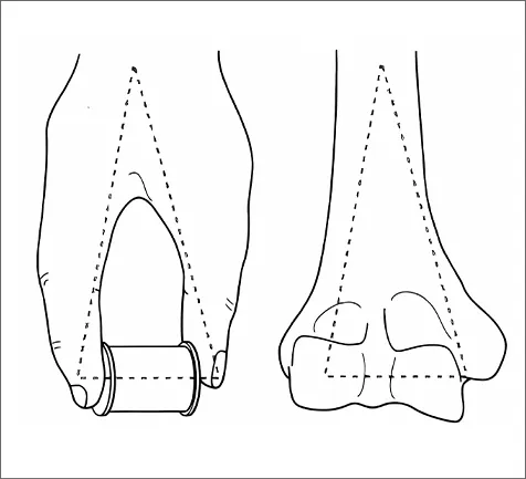
1. Anatomy of the Distal Humerus
The distal humerus consists of the medial and lateral columns, which include the epicondyles and condyles.
2. Mechanism of Injury
Distal humerus fractures are caused by direct trauma (e.g., falls) or indirect forces (e.g., twisting or muscle pull).
3. AO Classification
The AO classification divides distal humerus fractures into three main types: A , B , and C .
4. Surgical Treatment
Surgical treatment follows AO principles: anatomical reduction, stable fixation, and early rehabilitation.
5. Clinical Value
Locking plates offer superior biomechanical stability, particularly in osteoporotic bone.
6. CZMEDITECH Plate Models
CZMEDITECH offers three models: extraarticular (01.1107), lateral (5100-17), and medial (5100-18) plates.

Why is the distal humerus prone to fractures?
As a crucial part of the elbow joint, distal humerus fractures often result from "direct trauma" (such as a fall landing on the elbow) or "indirect trauma" (such as twisting or throwing actions).
- Muscle Pulling Forces
The medial column includes the medial part of the metaphysis of the humerus, the medial epicondyle, and the medial condyle, including the trochlea of the humerus.
·Strong contraction of internal rotator muscles
·Strong contraction of elbow flexor muscles
- High-Energy Trauma
External forces such as traffic accidents or falls from height can result in comminuted fractures or involve the articular surface.
Coronoid Fossa and Olecranon Fossa
·Traffic accidents
·Falls from height


Treatment Principles:
Following the AO philosophy: "Anatomical reduction, stable fixation, and early functional exercise."
High-Energy Trauma
External forces such as traffic accidents or falls from height can result in comminuted fractures or involve the articular surface.
Treatment Principles
Anatomical reduction
Stable fixation
Early functional exercise
Surgical Indications
Articular displacement >2mm
Open fractures
Combined neurovascular injury
Failure of conservative treatment
Plate Fixation Strategie
Dual Plate Technique
Suitable for type C fractures. Fixation from both medial (e.g., anatomical locking plate) and lateral (e.g., parallel plate) sides provides 3D stability and reduces the risk of postoperative rotational deformity.
Single Plate Technique
Used for type A and partial type B fractures. Pre-contoured plates conforming to distal humerus anatomy minimize soft tissue dissection.
Minimally Invasive Approach
Combined with percutaneous screw placement to reduce infection risk and preserve periosteal blood supply.
Biomechanical Advantage
Locking plates provide angular stability, especially beneficial for osteoporotic patients.
Functional Recovery Guarantee
Anatomical reduction preserves elbow joint mobility to the greatest extent, reducing complications such as nonunion or malunion.
Customized Design
Plates shaped for specific fracture types (e.g., intercondylar ridge support plates) optimize force transmission and accelerate bone healing.
Our distal humerus locking plate series is specially designed for complex distal humeral fractures. With anatomical contouring, locking screw technology, and multiple specifications, it offers safe, stable, and flexible fixation solutions for clinical surgery.


