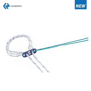
The Ultimate Guide to PEEK Anchors: Properties, Applications, and Advantages
If you're looking for a material that can provide superior strength, toughness, and chemical resistance, PEEK (polyetheretherketone) is the perfect choice. PEEK is a high-performance polymer that has become popular in many industries due to its outstanding mechanical properties, biocompatibility, and thermal stability. In this article, we'll discuss PEEK anchors, their properties, applications, and advantages.
What are PEEK Anchors?
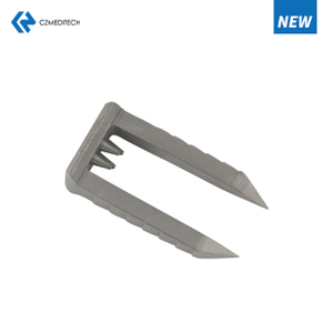
PEEK anchors are fasteners made of PEEK material that are designed to provide strong and reliable anchoring in various applications. They are commonly used in the medical, aerospace, automotive, and oil and gas industries, among others. PEEK anchors are known for their ability to withstand high loads, temperature changes, and harsh environments.
Properties of PEEK Anchors
PEEK anchors have many properties that make them superior to other materials. Some of the most notable properties of PEEK anchors include:
High Strength and Toughness
PEEK anchors have a high tensile strength and toughness, making them ideal for applications that require strong and reliable anchoring. They can withstand high loads and impact without breaking or deforming.
Chemical Resistance
PEEK anchors are highly resistant to many chemicals, including acids, bases, and organic solvents. This makes them ideal for applications where exposure to harsh chemicals is expected.
Thermal Stability
PEEK anchors have excellent thermal stability, withstanding high temperatures up to 300°C without melting or degrading. They are also resistant to cold temperatures, making them ideal for use in cryogenic applications.
Biocompatibility
PEEK anchors are biocompatible, making them safe for use in medical and dental applications. They are also non-toxic and non-carcinogenic, making them an environmentally friendly option.
Applications of PEEK Anchors
PEEK anchors have a wide range of applications, including:
Medical and Dental Applications
PEEK anchors are commonly used in medical and dental applications, such as orthopedic implants, dental implants, and spinal implants. Their biocompatibility and resistance to corrosion and wear make them ideal for use in the human body.
Aerospace Applications
PEEK anchors are used in the aerospace industry due to their high strength and resistance to temperature changes and chemicals. They are commonly used in aircraft engines, avionics, and structural components.
Automotive Applications
PEEK anchors are used in the automotive industry due to their resistance to wear, temperature changes, and chemicals. They are commonly used in engines, transmissions, and fuel systems.
Oil and Gas Applications
PEEK anchors are used in the oil and gas industry due to their resistance to high pressure, temperature changes, and chemicals. They are commonly used in downhole tools, valves, and pumps.
Advantages of PEEK Anchors
PEEK anchors offer many advantages over other materials, including:
Lightweight
PEEK anchors are lightweight compared to other metals, making them ideal for applications where weight is a concern.
High Strength-to-Weight Ratio
PEEK anchors have a high strength-to-weight ratio, making them ideal for applications where high strength is required but weight must be kept to a minimum.
Corrosion Resistance
PEEK anchors are highly resistant to corrosion, making them ideal for use in harsh environments.
Wear Resistance
PEEK anchors are highly resistant to wear, making them ideal for use in applications where abrasion is a concern.
Easy to Machine
PEEK anchors are easy to machine, making them a cost-effective option for many applications.
Conclusion
PEEK anchors are a versatile and high-performance fastener option for a wide range of applications. With their exceptional properties, applications, and advantages, they are becoming increasingly popular in many industries. From medical and dental implants to aerospace and automotive components, PEEK anchors provide reliable and strong anchoring with superior properties that make them stand out from other materials.
FAQs
What is PEEK?
PEEK stands for polyetheretherketone, which is a high-performance polymer known for its excellent mechanical properties, thermal stability, and biocompatibility.
What are PEEK anchors used for?
PEEK anchors are used for reliable and strong anchoring in various applications, such as medical implants, aerospace components, automotive parts, and oil and gas equipment.
What are the advantages of PEEK anchors?
PEEK anchors offer advantages such as high strength and toughness, chemical and thermal resistance, biocompatibility, lightweight, high strength-to-weight ratio, corrosion resistance, wear resistance, and easy machinability.
What are the applications of PEEK anchors?
PEEK anchors have applications in medical and dental implants, aerospace components, automotive parts, and oil and gas equipment.
Are PEEK anchors environmentally friendly?
Yes, PEEK anchors are environmentally friendly as they are non-toxic and non-carcinogenic, making them a safe and sustainable option for various applications.
English
Français
Русский
Español
العربية
Português
Deutsch
italiano
日本語
한국어
Nederlands
Tiếng Việt
ไทย
Polski
Türkçe
አማርኛ
ພາສາລາວ
ភាសាខ្មែរ
Bahasa Melayu
ဗမာစာ
தமிழ்
Filipino
Bahasa Indonesia
magyar
Română
Čeština
Монгол
қазақ
Српски
हिन्दी
فارسی
Kiswahili
Slovenčina
Slovenščina
Norsk
Svenska
українська
Ελληνικά
Suomi
Հայերեն
עברית
Latine
Dansk
اردو
Shqip
বাংলা
Hrvatski
Afrikaans
Gaeilge
Eesti keel
Māori
नेपाली
Oʻzbekcha
latviešu
অসমীয়া
Aymara
Azərbaycan dili
Bamanankan
Euskara
Беларуская мова
भोजपुरी
Bosanski
Български
Català
Cebuano
Corsu
ދިވެހި
डोग्रिड ने दी
Esperanto
Eʋegbe
Frysk
Galego
ქართული
guarani
ગુજરાતી
Kreyòl ayisyen
Hausa
ʻŌlelo Hawaiʻi
Hmoob
íslenska
Igbo
Ilocano
Basa Jawa
ಕನ್ನಡ
Kinyarwanda
गोंगेन हें नांव
Krio we dɛn kɔl Krio
Kurdî
Kurdî
Кыргызча
Lingala
Lietuvių
Oluganda
Lëtzebuergesch
Македонски
मैथिली
Malagasy
മലയാളം
Malti
मराठी
ꯃꯦꯇꯥꯏ (ꯃꯅꯤꯄꯨꯔꯤ) ꯴.
Mizo tawng
Chichewa
ଓଡ଼ିଆ
Afaan Oromoo
پښتو
ਪੰਜਾਬੀ
Runasimi
Gagana Samoa
संस्कृत
Gaelo Albannach
Sepeti
Sesotho
chiShona
سنڌي
Soomaali
Basa Sunda
Wikang Tagalog
Тоҷикӣ
Татарча
తెలుగు
ትግንያውያን
Xitsonga
Türkmençe
संस्कृत
ئۇيغۇرچە
Cymraeg
isiXhosa
ייִדיש
Yorùbá
isiZulu

